
Creating a comprehensive single cell spatial atlas of the human parathyroid gland
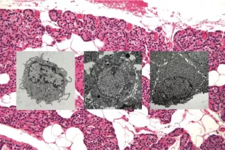
The parathyroid gland is comprised of multiple different cell types. By light microscopy (background) the differences can be subtle, but at the electron microscope level variations in cellular content, shape, and size are readily apparent (gray scale images).
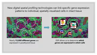
A precise understanding of the cellular composition and physical architecture of the human parathyroid gland is essential for the development of replacement therapies that can faithfully and sustainably reconstitute native gland function. Based on histological appearance, glandular architecture, and interrogative biochemical characterization of human parathyroid cells in situ, it is apparent that this complex endocrine organ is comprised of multiple, functionally distinct cell types. We seek to define the discrete cell types within human parathyroid tissue using single cell transcriptional profiling coupled with multiplexed spatial indexing. No such data have been generated for the human parathyroid gland to date. To address this major gap in knowledge, we are developing a comprehensive map of the parathyroid gland at single cell resolution where the physical location and spatial relationships between cells expressing distinct transcriptional programs can be visualized. First, we are performing a single cell RNAseq analysis of normal human organ donor parathyroid tissue to identify transcriptionally distinct cellular subpopulations within the gland. Second, we are localizing these individual cell types in sections of intact parathyroid tissue by highly multiplexed transcript and protein in situ quantitation, utilizing a Nanostring digital spatial profiling platform. These data will establish a critical reference standard defining the cellular constituents of the human parathyroid gland and determining the relative locations of the different cell types and their structural relationships within normal parathyroid tissue. This invaluable resource will be essential for guiding the development of replacement therapies and validating the fidelity of stem cell differentiation efforts to reconstitute native gland structure and function.
This work is supported by grants from the California Institute of Regenerative Medicine (CIRM 11437) and the Hypopara Foundation (Hypopara-21-001-01).
Multiple different cell types in the parathyroid gland work together to monitor and regulate blood calcium levels. Identifying and profiling these cell types at the molecular level is essential for understanding how to reconstitute normal calcium homeostasis in patients who have lost parathyroid gland function.
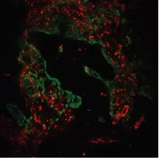
The parathyroid gland is physically organized into distinct structural compartments of unknown function.
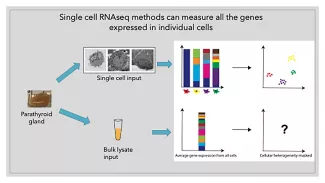
Newly developed single-cell profiling methods will allow us to define gene expression patterns that define each constituent cell type of the parathyroid gland and map their specific physical locations in the intact tissue.